I. Brightfield Transmitted Light Microscope
A. Light transmitted through specimen
interacts with specimen to create an image
| SPECIMEN PROPERTIES | EFFECTS ON IMAGE LIGHT |
| Different degrees of opacity, Large differences in refractive index | Brightness contrast |
| Interference or absorption colors | Color contrast |
| Different thickness, small differences in refractive index | Phase changes |
B. Thick specimens need to be be
sectioned
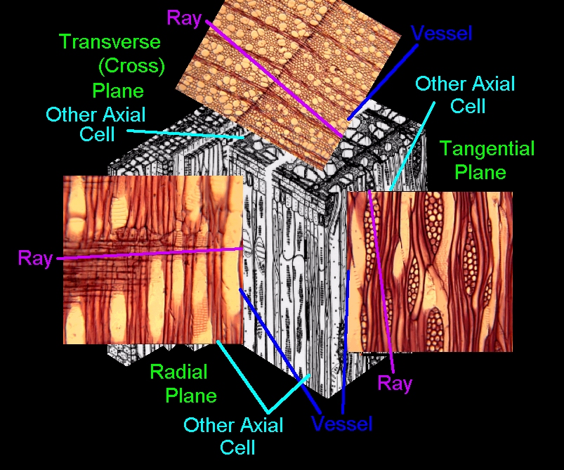
| TECHNIQUE | MOST OFTEN USED FOR | SECTION THICKNESS RANGE (micrometers) | IMAGE | |||
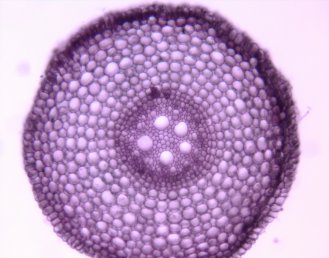
| Free hand sections | Quick for fresh herbacious material and soft wood | 10 - 40 | Zea Root | |||
| Vibratome | Fresh herbacious material and soft wood | 5 - 40 | Pinus Leaf | |||
| Sliding microtome | Harder wood | 1 - 40 | Wood Sections | |||
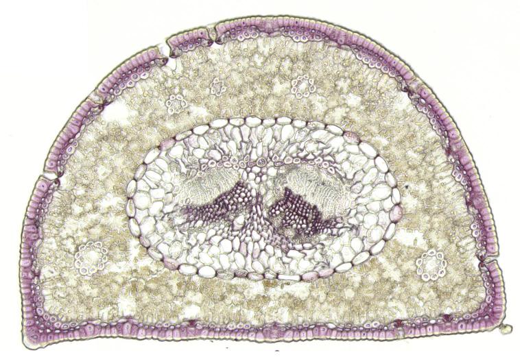
| Rotary microtome | Paraffin or soft plastic embedded material | 1 - 50 | Coleus Shoot Apex | |||
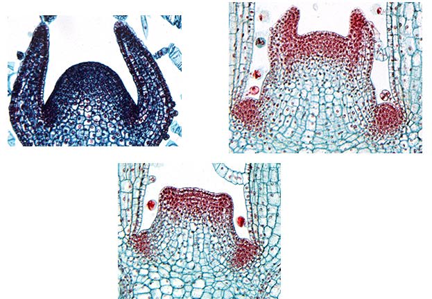
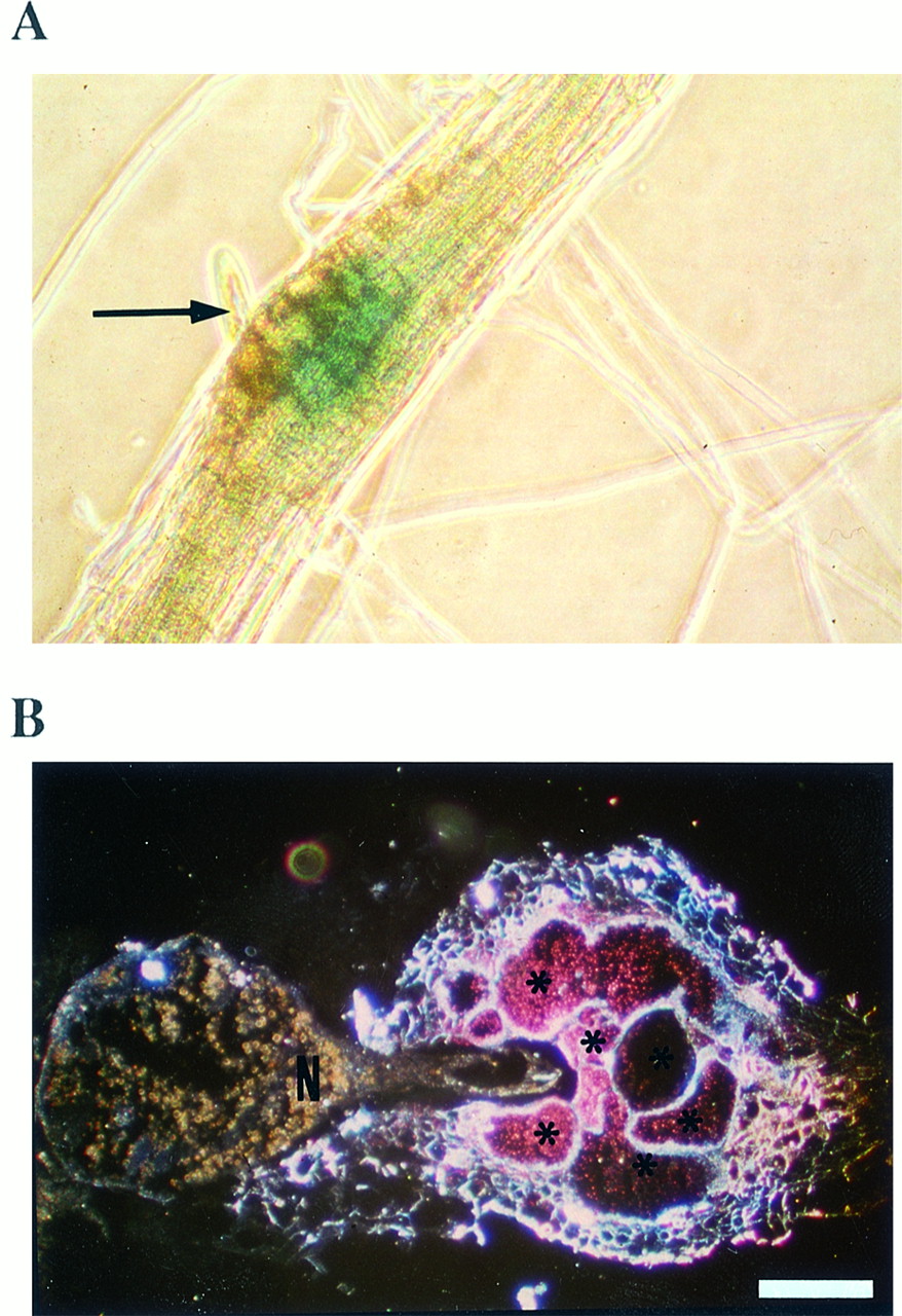
| Cryomicrotome | Frozen material | 1 - 50 | Arabipopsis root nematode gall | |||
| Ultramicrotome | Hard plastic embedded material |
|
Meristematic Cells |
C. Adjustable parameters
1. Magnification
2. Resolution
3. Contrast
a. Introduction of general or specific dyes
b. Modification of microscope
A. Useful for observing fresh material with little inherent contrastB. Condenser with a circular aperture called the phase annulus
C. Objective with a circular ring of phase retarding material called the phase ring
D. Light passing through specimen is shifted in phase compared to background light passing through mounting medium
E. Specimen light either adds to or subtracts from background light generating lighter or darker regions of contrast
F. Contrast depends on differences in refractive indices of specimen and mounting medium
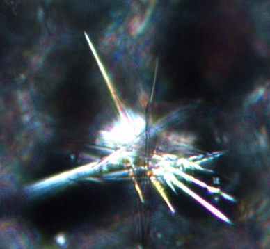
III. Polarized
Light Microscope
A. Useful for observing crystaline or quasi-crystaline structureB. Below the condenser is a piece of polarizing material called the polarizer
C. Between the objective and the ocular is another piece of polarized material called the analyzer
D. Light passing through condenser is polarized into a single plane
E. If the analyzer is rotated 90o relative to polarizer, no light reaches the ocular and the image is black = crossed polarized light
F. If specimen has crystals or quasi-crystaline structure (starch and cellulose) then polarized light that passes through these objects will be rotated at some angle relative to the initial plane of polarization.
G. This rotated light is called birefringence and if rotated enough will pass through the analyzer and be visible.
H. The analyzer can be rotated relative to the polarizer to examine birefringence that results from different angles of rotation.
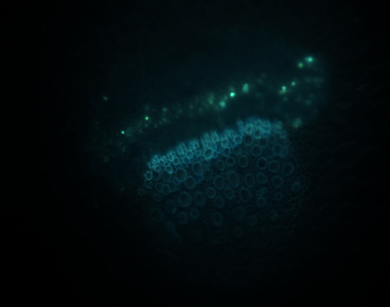
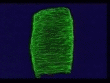
A. Useful for observing fluorescent material1. Fluorescence results when material absorbs light of one wavelength and emits light of a longer wavelength (lower energy)
2. Different materials absorb and fluoresce different wavelengths of lightB. Laser beam is shined on specimen through the objective
C. Depending on the material, fluorescence may take place and be collected by the objective
D. Depth of field is severly restricted so that only fluorescence coming from a very thin plane in the section is collected
1. This results in a very sharp image since no background light (glare) is collected
E. Image capture and fine focus of microscope controlled by a computer
1. The computer creates a digital frame of the restricted depth of field image
2. The computer then moves the specimen to a different focal plane and captures a new image
3. After scanning through a range of foci, the computer combines all the image framesF. Final image is very clean (high signal to noise ratio) since it contains very little noise from out of focus parts of the specimen
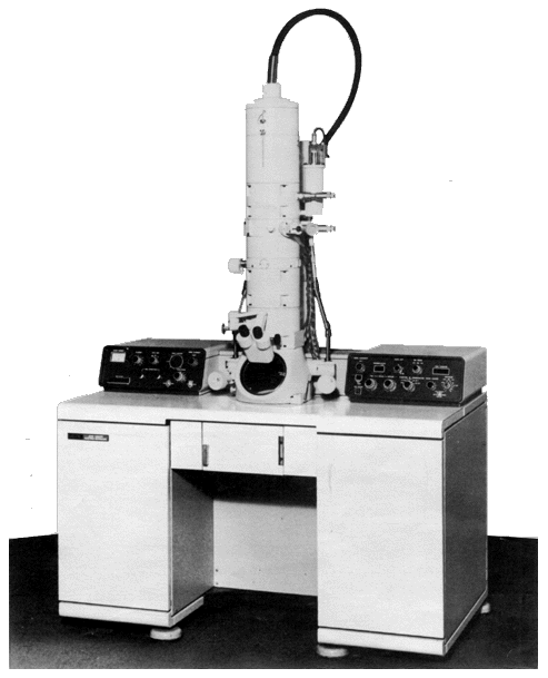
V. Transmission
Electron Microscope
A. Useful for resolving objects below the resolution of lightVI. Scanning Electron MicroscopeB. Design is basically like compound light microscope but TEM
1. Uses electrons focused by electromagnetic lenses in a vacuum environment
2. Specimens are much thinner
3. Specimens stained with heavy metals since most biological material is transparent to electrons
4. Image can't be seen directly, but rather is viewed via a fluorescent screen
5. Images are gray scale since electrons have no color
A. Useful for observing surface detailsB. Electron beam is focused on surface of specimen with electromagnetic lenses in a vacuum
C. Beam scan back and forth across specimen in syncrony with the electron beam of a cathode ray tube
D. Electrons of beam cause various kinds of signals to be released by atoms in specimen
1. Secondary electrons
2. Backscattered electrons
3. Photons
4. X-raysE. The intensity of these signals varies in the specimen and on the display screen creating the image
| MICROSCOPE | MAGNIFICATION RANGE | RESOLUTION | DEPTH OF FIELD |
| LIGHT | 1 - 1,000 X | 0.2 um | 2 um |
| TRANSMISSION ELECTRON | 2,000 - 900,000 X | 0.21 - 0.5 nm | 0.11 um |
| SCANNING ELECTRON | 2 - 300,000 X | 3.5 - 6 nm | up to 1 cm |